Abstract
Introduction
Central venous cannulation is widely used in critically ill patients. Although the landmark-guided technique has a high success rate, it can cause serious complications due to anatomical variations. The aim of this study was to determine the anatomical variations between the internal jugular vein (IJV) and carotid artery (CA). Another aim of the study is to reveal the risk of complications associated with cannulation performed using anatomical landmark guidance.
Methods
This retrospective study included 61 patients admitted to the pediatric intensive care unit. Information was obtained from the recorded files. Descriptive statistical analyses, the chi-square, Fisher’s exact, and Bonferroni tests, and multivariate logistic regression analysis were used as statistical tools.
Results
Thirty-six patients were male and 25 were female. The most common admission diagnosis was respiratory failure (60.1%). The mean age of the patients was 58.1±62 months (2 to 192 months). Most catheterizations were performed unilaterally. The right side was predominantly preferred. The most common locations of the IJV and CA on both sides were anterolateral. Significant overlap was detected on the right side in 18 patients (29.5%) and on the left side in 21 patients (34.4%). It was also observed that, on the left side, as weight and age decreased, the overlap increased significantly.
Conclusion
Our findings support the routine use of ultrasound (USG)-guided catheterization in children. This study has shown that the risk of complications is high in catheterization procedures performed without USG, especially in infants and underweight children.
Keywords: Anatomical variations, carotid artery, internal jugular vein, pediatric, USG-guided catheterization
Introduction
Central venous cannulation (CVC) is a widely used procedure by pediatric intensivists, surgeons, and anesthesiologists. This procedure is used for various indications, such as blood sampling, vasopressor administration, parenteral nutrition, antibiotic administration and long-term chemotherapy, hemodialysis, and plasmapheresis.- The internal jugular vein (IJV), femoral, and subclavian veins are commonly used for this purpose in children. The IJV is preferred in children due to its anatomic position, accessibility, and low complication rates. A right-sided approach is generally recommended for CVC insertion because the right IJV has a larger diameter and lies more superficially.
Although ultrasound (USG)-guided IJV cannulation procedures are the gold standard, CVC is still performed using anatomical landmark guidance and is associated with variable success and complication rates in developing countries such as our country. The Sedillot triangle has been used for IJV punctures for years as an anatomical landmark. In this anatomical landmark guidance technique, it is assumed that the IJV is usually lateral to the carotid artery (CA)., Although cannulation procedures using this technique generally have a high success rate, adverse events may occur at varying rates depending on multiple factors., During cannulation, accidental arterial puncture, hematoma, or both can decrease success rates. However, USG-guided cannulation will increase the success of the procedure and reduce the likelihood of complications. Knowledge of the variable anatomical relationship between the CA and the IJV will minimize undesirable complications during the procedure. Limited information on this topic is available for children. Therefore, we analyze the anatomical relationship between CA and IJV in pediatric cases by point-of-care USG.
The objective of this study was to determine the anatomical variations between the IJV and the CA. Another aim of the study is to reveal the risk of possible complications in cannulations using anatomical landmark guidance.
Materials and Methods
This study, which retrospective analysis of prospectively collected data, in cluded 61 patients who were admitted to the Pediatric Intensive Care Unit of Gaziantep University Gaziantep Research and Training Hospital between 01.07.2023 and 01.03.2024, aged between 28 days and 18 years, and underwent IJV catheterization using USG imaging. Exclusion criteria for the study were known abnormal neck anatomy, neck trauma, neck tumor, previous neck surgery, or previous IJV catheterization.
Demographic data (age, gender, weight), clinical characteristics (mortality scores, main hospitalization diagnoses, co-morbid conditions, indication for CVC), and catheterization procedure information (side where the catheter is inserted, artery-vein relationship, and number of catheters inserted) were obtained from the recorded files.
The USG examination was performed in the supine position. The examination was performed with the neck extended and the head rotated 15-30° to the opposite side, as adopted for IJV cannulation. A small rolled towel was placed under the child’s shoulder to ensure neck extension. The same pediatric intensivist performed all USG examinations. This pediatric intensivist had been performing CVC cannulation on patients for over 13 years. For the last 10 years, he has also been performing USG-guided CVCs. A portable USG “(ESAOTE/MYLAB 30CV, ESAOTE S.p.A. Florence/Italy)” machine with a linear array (vascular) probe was used.
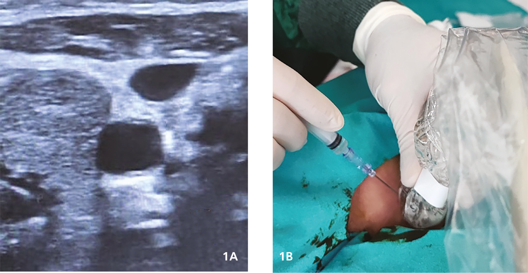
Sedillot’s triangle is defined as a triangle bounded by the medial and lateral heads of the sternocleidomastoid muscle, with its base formed by the medial end of the clavicle. The probe was placed perpendicular to the apex of the Sedillot triangle (). The apex of the triangle was the point where a needle was inserted into the IJV. Vessels were visualized in transverse section using B-mode and color Doppler (). The patients were sedated (fentanyl 1-2 μg/kg and/or midazolam 0.15 mg/kg). Afterward, the catheter was introduced using the Seldinger technique under sterile conditions.
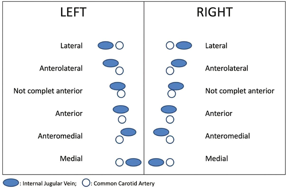
The relationship between CA and IJV was classified according to the degree to which the IJV overlapped the CA (), consistent with previous studies., , This classification is based on the locations of the IJV and CA and on their overlap rates. The lateral position is defined as the IJV being in a lateral location and not overlapping with the CA; the anterolateral position is defined as the IJV being in a lateral location and having <50% overlap with the CA; the incomplete anterior position is defined as the IJV being in a lateral location and having >50-99% overlap with the CA; the anterior position is defined as the IJV completely overlapping the CA; the anteromedial position is defined as the IJV being in a medial location and having <50% overlap with the CA; and the medial position is defined as the IJV being in a medial location and not overlapping with the CA. When ultrasonography was not used, positions at risk of CA puncture during IJV puncture (anterior and incomplete anterior) were considered “significant overlap”. The l ateral position (no risk of CA puncture) and the anterolateral position (low risk of CA puncture) were considered “non-significant overlap”.
Ethical approval for this study was provided by the Gaziantep University Non-Invasive Clinical Research Ethics Committee (approval no: 2024/89, date: 27.03.2024). Informed consent was waived for this research due to the retrospective nature of the studies.
Statistical Analysis
All data were analyzed using the Statistical Package for the Social Sciences (SPSS) version 22.0 (SPSS Inc., Chicago, IL, USA). Descriptive statistical analyses were performed for demographic characteristics. The results were reported as mean ± standard deviation. Categorical variables were compared using the chi-square, Fisher’s exact, and were expressed as counts and percentages. Multivariate logistic regression was performed to identify independent risk factors associated with significant overlap. P<0.05 was considered statistically significant.
Results
A total of 61 patients were included in the study. Of the patients, 36 were male and 25 were female. The most common admission diagnoses were respiratory failure (60.1%), neurological diseases (40.1%), and sepsis (32.8). Forty-one (67.2%) patients had comorbidities. The mean age was 58.1±62 months (2 to 192 months), and the mean weight was 17.7±15 kg (4 to 70 kg). Clinical and demographic characteristics are presented in .
Table 1. Clinical and demographic characteristics of the patients
Parameters | n | % |
|---|---|---|
Gender | ||
Male | 36 | 59.1 |
Female | 25 | 40.9 |
Admission diagnoses* | ||
Respiratory failure | 37 | 60.1 |
Neurological diseases** | 25 | 40.1 |
Sepsis | 20 | 32.8 |
Congenital heart diseases | 7 | 11.5 |
Oncological diseases | 7 | 11.5 |
Metabolic diseases | 7 | 11.5 |
Trauma | 5 | 8.2 |
Post-arrest | 4 | 6.6 |
Hepatic failure | 4 | 6.6 |
Renal failure | 4 | 6.6 |
Others | 4 | 6.6 |
Co-morbidities | ||
Yes | 41 | 67.2 |
No | 20 | 32.8 |
Continuous data | Mean ± SD | Min-max |
Age (months) | 58.1±62 | 2-192 |
Weight (kg) | 17.7±15 kg | 4-70 |
*: There may be several diagnoses, **: Epileptic seizures, encephalitis, intracranial hemorrhage, altered consciousness, SD: Standard deviation
Mostly unilateral catheterization was performed. The right side was mostly preferred. Blood sampling, sedoanalgesia, and the need for inotropes were the most common indications for CVC. The most common IJV and CA locations on both sides were anterolateral. Significant overlap was detected in 18 (29.5%) patients on the right side and 21 (34.4%) patients on the left side. Details of CVC procedures are given in .
Table 2. Details of CVC procedures
Parameters | n | % |
|---|---|---|
CVC procedures | ||
Unilaterally | 57 | 93.5 |
Bilaterally | 4 | 6.5 |
CVC side* | ||
Right | 47 | 77.1 |
Left | 18 | 29.5 |
CVC indications | ||
Blood sampling | 61 | 100 |
Sedoanalgesia | 35 | 57.4 |
Need for inotropes | 34 | 55.7 |
Dialysis | 14 | 22.9 |
Multiple antibiotic therapy | 5 | 8.2 |
TPN | 4 | 6.5 |
Chemotherapy | 2 | 3.3 |
IJV and CA locations and overlap rates | ||
Right side | ||
Lateral | 20 | 32.8 |
Anterolateral | 23 | 37.7 |
Incomplet anterior | 13 | 21.3 |
Anterior | 5 | 8.2 |
Anteromedial | 0 | 0 |
Medial | 0 | 0 |
Left side | ||
Lateral | 18 | 29.5 |
Anterolateral | 22 | 36.1 |
Incomplet anterior | 13 | 21.3 |
Anterior | 8 | 13.1 |
Anteromedial | 0 | 0 |
Medial | 0 | 0 |
Significant overlap | ||
Right side | 18 | 29.5 |
Left side | 21 | 34.4 |
*: Bilateral catheterization was performed in 4 patients, CVC: Central venous cannulation, TPN: Total parenteral nutrition, IJV: Internal jugular vein, CA: Carotid artery
Factors that may be related to significant overlap were examined (). On the right side, a high rate of significant overlap was observed among children weighing less than 10 kg. However, this difference was not statistically significant (p=0.065). On the left side, overlap increased significantly as weight and age decreased. Multivariate logistic regression analysis revealed that gender and weight were independent risk factors for significant overlap ().
Table 3. Parameters that may be associated with significant overlap on the right side
Parameters | Significant overlap (+), n=18 | Significant overlap (-), n=43 | p-value | ||
|---|---|---|---|---|---|
n | % | n | % | ||
Gender | 0.265 | ||||
Male | 13 | 72.2 | 24 | 55.8 | |
Female | 5 | 27.8 | 19 | 44.2 | |
Co-morbidities | 0.395 | ||||
Yes | 11 | 61.1 | 30 | 69.8 | |
No | 7 | 38.9 | 13 | 30.2 | |
Age (months) | 0.259 | ||||
1-36 | 12 | 66.7 | 22 | 51.2 | |
37-72 | 2 | 11.1 | 3 | 7.0 | |
73-144 | 4 | 22.2 | 11 | 25.6 | |
145-216 | 0 | 0 | 7 | 16.3 | |
Weight (kg) | 0.065 | ||||
1-10 | 14 | 77.8 | 19 | 44.2 | |
11-30 | 3 | 16.7 | 11 | 25.6 | |
31-60 | 1 | 5.5 | 12 | 27.9 | |
60-90 | 0 | 0 | 1 | 2.3 | |
Table 4. Parameters that may be associated with significant overlap on the left side
Parameters | Significant overlap (+), n=21 | Significant overlap (-), n=40 | p-value | ||
|---|---|---|---|---|---|
n | % | n | % | ||
Gender | 0.275 | ||||
Male | 15 | 71.4 | 22 | 55.0 | |
Female | 6 | 28.6 | 18 | 45.0 | |
Co-morbidities | 0.056 | ||||
Yes | 11 | 52.4 | 30 | 75.0 | |
No | 10 | 47.6 | 10 | 25.0 | |
Age (months) | 0.038* | ||||
1-36 | 15 | 71.4 | 19 | 47.5 | |
37-72 | 3 | 14.3 | 2 | 5.0 | |
73-144 | 3 | 14.3 | 12 | 30.0 | |
145-216 | 0 | 0 | 7 | 17.5 | |
Weight (kg) | 0.029* | ||||
1-10 | 15 | 71.4 | 16 | 40.0 | |
11-30 | 4 | 19.1 | 11 | 27.5 | |
31-60 | 2 | 9.5 | 12 | 30.0 | |
60-90 | 0 | 0 | 1 | 2.5 | |
Table 5. Factors related to significant overlap were evaluated using multivariate logistic regression analysis (parameters that were significant are given)
Parameters | Odds ratio | 95% CI | p-value |
|---|---|---|---|
Right side | |||
Weight | 1.376 | 3.959 (1.587-9.877) | 0.003 |
Gender (male) | 1.327 | 3.769 (1.004-14.154) | 0.049 |
Left side | |||
Weight | 1.326 | 3.768 (1.618-8.772) | 0.002 |
Gender (male) | 1.254 | 3.504 (1.009-12.164) | 0.048 |
CI: Confidence interval
Discussion
The use of IJV for CVC is widely preferred by physicians, especially intensivists and anesthesiologists. Many, complications such as inadvertent arterial puncture, hematoma, accidental intraarterial cannulation, and IJV-CA fistula, may develop during this procedure. To minimize these complications, IJV-CA locations must be well-known. Although the use of USG during the CVC procedure is recommended, it is not a routine practice. This study highlights complications that may develop from the anatomical relationship between the CA and the IJV when USG is not used.
Many studies have shown that the right IJV is preferred for CVC procedures. Turba et al. reported that cannulation of the IJV was successfully achieved in the 184 patients with a patent IJV, 75% on the right. Another study stated that the safe position for CVC is 85% on the right and 72% on the left. Maneenai and Arjhansiri reported that the right IJVs were usually larger than the left IJVs. Additionally, one study reported that the right IJV was wider than the left. In our study, consistent with previous studies, the right IJV was preferred in 77.1% of cases.
The relationship between CA and IJV has been investigated using different methods in numerous adult studies. In a study of adult patients evaluated by CT, the IJV was predominantly in the lateral position (94.2%). A study found that the IJV was located mostly in the anterolateral position (81% on the right side and 71% on the left side).Another adult study reported that, in the neutral position, the anterolateral position was most common on both the right and left sides whereas when the head was turned to the other side, the anterior position was more common on both sides. Lorchirachoonkul et al. reported that in all positions where the head was turned to the right and left at certain angles, the IJV was mostly in the lateral position. In one of a limited number of pediatric studies, the right IJV was located laterally in 84% of cases. Our results are similar to those of Parmar et al.The primary reason our findings differ from the existing literature is that the age group evaluated is different. In the pediatric study discussed, only children weighing less than 4000 g were evaluated; therefore, differences compared with our results may have occurred.
Many previous studies have examined the relationship between IJV, CA, and vertebral artery (VA). The common goal of these studies is to identify high-risk vascular locations and significant rates of overlap. In a study evaluating preschool children, more than half were at moderate to high risk for VA puncture at the time of IJV cannulation. In a study of adults, the rate of significant overlap was 5.8%. In an adult study examining the relationship between IJV and CA, 15% of patients had a dangerous position on the right side, and 28% had a dangerous position on the left. In an ultrasonography study, significant overlap was 30.7% on the right and 41.1% on the left in the neutral position; when the head was turned to the opposite side, it was 37.3% on the right and 46.6% on the left. In our study, significant overlap rates were examined using USG with the head turned to the opposite side. In this case, we observed a significant overlap of 29.5% on the right and 34.4% on the left. Additionally, this significant left-sided overlap increased with decreasing age and weight. These rates indicate a high risk associated with catheterization performed without USG guidance in pediatric patients, especially infants.
Study Limitations
The most important limitation of the study is that, while the relationship between IJV and CA was evaluated, the relationship with VA was not. Another limitation of the study is the relatively small number of cases evaluated at a single center. We would like to state that the sample size may reduce the statistical power of the study, and that subgroup findings should be interpreted cautiously.
In addition, if adults were evaluated alongside children, the effect of age on the relationship between IJV and CA could be understood more clearly.
Conclusion
Despite its limitations, we believe that our study will contribute to the literature. Our findings support the routine use of USG-guided catheterization in children. This study has shown that the risk of complications will be high in catheterization procedures performed without using USG, especially in infants and underweight children.
Footnotes
Ethics
Ethics Committee Approval: Ethical approval for this study was provided by the Gaziantep University Non-Invasive Clinical Research Ethics Committee (approval no: 2024/89, date: 27.03.2024).
Informed Consent: Informed consent was waived for this research due to the retrospective nature of the studies.
Acknowledgments
We would like to thank our clinic head, Prof. MD., Mehmet Keskin, for his contributions to the realization of our study.
Authorship Contributions
Surgical and Medical Practises: Ç.K., M.B., Concept: Ç.K., M.B., Design: Ç.K., M.B., Data Collection or Processing: O.D., Analysis or Interpretation: Ç.K., M.B., Literature Search: Ç.K., M.B., O.D., Writing: Ç.K.
Conflict of Interest: No conflict of interest was declared by the authors.
Financial Disclosure: The authors declared that this study received no financial support.
References
So KY, Kim SH, Kim DW. Anatomical relationship of the internal jugular vein and the common carotid artery in Korean: a computed tomographic evaluation. Anesth Pain Med. 2015;10:118-23.
Eisen LA, Narasimhan M, Berger JS, Mayo PH, Rosen MJ, et al. Mechanical complications of central venous catheters. J Intensive Care Med. 2006;21:40-6.
Wu SY, Ling Q, Cao LH, Wang J, Xu MX, et al. Real-time two-dimensional ultrasound guidance for central venous cannulation: a meta-analysis. Anesthesiology. 2013;118:361-75.
Parmar S, Parikh S, Mehta H. Anatomical variations of the internal jugular vein in relation to carotid artery: an ultrasound study. Int J Med Sci Public Health. 2013;2:223-8.
Maneenai N, Arjhansiri K. CT evaluation of anatomical variations of the internal jugular veins in Thai adults. Asian Biomedicine. 2013;7;803-11.
Umaña M, García A, Bustamante L, Castillo JL, Sebastián Martínez J. Variations in the anatomical relationship between the common carotid artery and the internal jugular vein: an ultrasonographic study. Colomb Med (Cali). 2015;46:54-9.
McGee DC, Gould MK. Preventing complications of central venous catheterization. N Engl J Med. 2003;348:1123-33.
Asouhidou I, Natsis K, Asteri T, Sountoulides P, Vlasis K, et al. Anatomical variation of left internal jugular vein: clinical significance for an anaesthesiologist. Eur J Anaesthesiol. 2008;25:314-8.
Turba UC, Uflacker R, Hannegan C, Selby JB. Anatomic relationship of the internal jugular vein and the common carotid artery applied to percutaneous transjugular procedures. Cardiovasc Intervent Radiol. 2005;28:303-6.
Ruesch S, Walder B, Tramèr MR. Complications of central venous catheters: internal jugular versus subclavian access--a systematic review. Crit Care Med. 2002;30:454-60.
Hutchinson J, Hor KF, Wong YM, Mitchell C. View of the internal jugular vein and carotid artery in their true anatomical position. Eur J Anaesthesiol. 2013;30:258-9.
Lau CS, Chamberlain RS. Ultrasound-guided central venous catheter placement increases success rates in pediatric patients: a meta-analysis. Pediatr Res. 2016;80:178-84.
Maecken T, Marcon C, Bomas S, Zenz M, Grau T. Relationship of the internal jugular vein to the common carotid artery: implications for ultrasound-guided vascular access. Eur J Anaesthesiol. 2011;28:351-5.
Hong JY, Koo BN, Kim WO, Choi E, Kil HK. Effect of head rotation on overlap and relative position of internal jugular vein to carotid artery in infants and children: a study of the anatomy using ultrasonography. J Crit Care. 2010;25:360.e9-360.13.
Domino KB, Bowdle TA, Posner KL, Spitellie PH, Lee LA, et al. Injuries and liability related to central vascular catheters: a closed claims analysis. Anesthesiology. 2004;100:1411-8.
Uzumcugil F, Yilbas AA, Akca B. Ultrasound-guided anatomical evaluation and percutaneous cannulation of the right internal jugular vein in infants <4000 g. J Vasc Access. 2020;21:92-7.
Lorchirachoonkul T, Ti LK, Manohara S, Lye ST, Tan SA, et al. Anatomical variations of the internal jugular vein: implications for successful cannulation and risk of carotid artery puncture. Singapore Med J. 2012;53:325-8.
Ganesh A, Paarel JP, Punnen GE, Siddarth R, Prakash KS, et al. Anatomical relationship between the internal jugular vein and the vertebral artery: an observational study in pre-school children using ultrasound imaging. J Anaesthesiol Clin Pharmacol. 2022;38:544-7.