Abstract
Introduction
Recently, some centers have used extubation during extracorporeal membrane oxygenation (ECMO) to eliminate barotrauma and volutrauma as a lung rest strategy. This study aims to demonstrate the use of extubation during ECMO in children.
Methods
This retrospective study was conducted from January 1, 2015, to April 1, 2023, in our pediatric intensive care unit.
Results
In this study, we presented six cases that were extubated during ECMO support. In addition, we followed 130 pediatric patients on ECMO in the same period. Two patients were primarily diagnosed with cardiomyopathy, one with myocarditis, two with congenital heart defect, and one with necrotizing pneumonia. The median age of patients was 99 (interquartile range 25-75) (16.5-192) months, and all were male. Venoarterial ECMO was connected to 4 patients, and venovenous ECMO was connected to 2 patients. Six patients were extubated during ECMO on the 5th, 12th, 3rd, 4th, 3rd and 14th days of their treatment, respectively. While the patients were extubated, three of them were supported by biphasic positive airway pressure, one was supported by nasal continuous positive airway pressure, and two were provided with supplementary oxygen. Three patients were extubated under ECMO and discharged.
Conclusion
The risk of mechanical ventilation related complications such as volutrauma and barotrauma could minimized in patients extubated under ECMO. In addition, sedatives, analgesics, and muscle relaxant related complications such as delirium, muscle weakness, or prolonged ventilation could reduced via awake ECMO.
Keywords: Extracorporeal membrane oxygenation, extubation, mechanical ventilation, child
Introduction
Extracorporeal membrane oxygenation (ECMO) is a widely used life-saving intervention for children with circulatory and respiratory failure that is refractory to standard therapies. Due to advances in extracorporeal life support (i.e., technical devices, cannulation technique, specialized ECMO teams), ECMO has become a safe tool in pediatric intensive care. These developments in ECMO may be used for pediatric and adult patients for weeks for recovery and bridge therapy to left ventricular assist device implantation or transplantation. ECMO performs this by regulating cellular respiration, improving tissue oxygenation, and allowing CO2 removal. It is, however, an invasive treatment method and can cause potential serious complications. ECMO has been considered the last treatment option to be used only when traditional treatments do not respond and only for a sufficient duration until the patient may be returned to more “standard” support modalities (mechanical ventilation, inotropic support, and cardiopulmonary resuscitation).
During ECMO, patients are often prescribed a lot of sedatives-analgesics and invasive mechanical ventilation (MV) to avoid fatal decannulation and facilitate ECMO performance. Sedation also reduces oxygen consumption, thereby improving arterial oxygen transport and organ perfusion. Nevertheless, both sedation and MV may cause complications. Long-term sedation and neuromuscular blockade therapy may cause muscle weakness, disability, tolerance, delirium, or prolonged recovery time. Possible invasive MV-related complications include tracheal irritation due to the intubation tube, discomfort, and the need for more sedation. In addition, it might cause ventilator-related complications, such as ventilator-associated pneumonia, pneumothorax, or respiratory muscle weakness. There is a growing interest in reducing exposure to narcotics, sedatives, and neuromuscular blocking agents and keeping ECMO patients awake and mobile to maintain muscle strength and shorten recovery times. Recently, a method of extubation from MV under ECMO, especially for patients with respiratory failure and as a bridge therapy to lung transplantation, has emerged.- However, extubation practices and management options during ECMO in pediatric intensive care units have not been adequately reported. This study presents the clinical findings of six children who were electively extubated during ECMO, along with an expanded discussion of two representative cases.
Materials and Methods
Patients
This retrospective study was conducted from 2015 to 2023 in our pediatric intensive care unit. We present six cases of extubation during ECMO support. In addition, we followed up 130 pediatric patients on ECMO during the same period. Patient demographic data and clinical and laboratory data were obtained from medical records.
In our clinic, 130 pediatric patients have been connected to ECMO so far, and 6 of them have been extubated during ECMO. Based on the extubation protocol in our clinic;
1. The patient’s FiO2<40% and SpO2>92% in IMV,
2. No respiratory effort at low pressures and minimal settings in the IMV,3. The patient becomes conscious after discontinuing sedation,4. After monitoring the IMV in continuous positive airway pressure (CPAP)/pressure support ventilation (PSV) mode (PSV 6, positive end-expiratory pressure 6 FiO2 40%) for 1-2 hours, there is no respiratory effort, and the patient is extubated if there is no acidosis or hypercarbia in the blood gas.
Ethical Considerations
Voluntary consent was obtained from the families of the patients included in the study. The study was approved by the Institutional Review Board of the Ankara University Faculty of Medicine (approval number: İ06-384-23, 2023/06).
Statistical Analysis
Statistical analyses were performed using SPSSv26.0 (Statistical Package for Social Sciences for MacOS, Inc., USA). Descriptive variables are presented as frequencies and proportions (%). Continuous variables are presented as median (lower-upper limit) values.
Results
Six children were extubated during ECMO support. The median age of patients was 99 [interquartile range (IQR) 25-75] (16.5-192) months, and all were male. Two patients were primarily diagnosed with cardiomyopathy, one with myocarditis, two with congenital heart defects, and one with necrotizing pneumonia. Venoarterial (VA) ECMO was connected to 4 patients, and venovenous-venoarteriovenous (VV) ECMO were connected to 2 patients. The peripheral ECMO was connected in five patients, whereas the central ECMO was connected in one patient. One patient who was connected to VV ECMO subsequently underwent venoarteriovenous (VAV) ECMO due to cardiogenic shock. Five patients were connected to ECMO for cardiac failure, and one patient was connected to respiratory failure.
Six patients were extubated during ECMO on the 5th, 12th, 3rd, 4th, 3rd and 14th days of treatment, respectively. The median number of days extubated during ECMO was 4.5 (IQR3-12.5) days. The durations of remaining extubation while on ECMO were 8, 9, 3, 6, 13, and 3 days for six patients, respectively. The median day of extubation duration under ECMO was 7(IQR 3-10) days. While the patients were extubated, three received biphasic positive airway pressure (BIPAP), one received nasal-CPAP (N-CPAP), and two were provided with supplementary oxygen. Five patients were reintubate during ECMO. Four patients were intubated due to increased respiratory distress, and one patient was intubated after decannulation. ECMO complications were observed in 4 patients, including bleeding in 4 cases, limb ischemia in 1 case, and circuit clotting in 3 cases. Three patients who were extubated under ECMO survived. Two patients were discharged with no neurological sequel.
Case 1
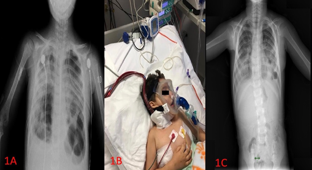
A 3-year-old male patient was transferred to our pediatric intensive care unit (PICU) following video-assisted thoracoscopic surgery (VATS) for necrotizing pneumonia and empyema. When the patient was admitted to intensive care, chest tubes were placed in both hemithorax. The chest X-ray taken in another center revealed pleural effusion, and the fluid was exudated via thoracentesis (). It was reported that the patient had cavitary lesions and consolidation areas that were evaluated in favor of bilateral necrotizing pneumonia and bilateral pleural effusion on CT thorax. The patient had respiratory distress and desaturation, requiring intubation. VATS was performed on the 2nd day of hospitalization. The patient was diagnosed with necrotizing pneumonia and metapneumovirus and was intubated following video-assisted thoracoscopic surgery (VATS). Oseltamivir was administered after the pleural fluid sample tested positive for influenza A (H1N1) and bocavirus via polymerase chain reaction (PCR). In addition, meropenem, vancomycin, and levofloxacin were initiated. On the 7th day of PICU admission, the patient was admitted to venovenous (VV) ECMO because of severe hypoxemia and respiratory acidosis in a high ventilator setting. The ECMO settings were an RPM of 4500/min and LPM of 1200 mL/min. Cardiac arrest occurred twice, and cardiopulmonary (CPR) resuscitation was performed within 5 and 12 min, respectively. The patient was extubated on the 12th day of ECMO, and BIPAP was subsequently applied at the full-face interface (, ). He was decannulate on the 22nd day of ECMO, but BIPAP was continued until two days after ECMO removal. The patient was subsequently transferred to the pediatric infection disease unit on the 38th day of PICU admission with normal vital signs. He was discharged on the 44th day of hospitalization.
PICU: Pediatric intensive care unit, ECMO: Extracorporeal membrane oxygenation
Case 2
A 17-month-old male patient was admitted to our PICU after the establishment of central VA ECMO due to fulminant myocarditis. The patient’s cardiac function was poor, with an ejection fraction of the left ventricular of 15%, and there was severe metabolic acidosis and recurrent ventricular tachycardia. ECMO was connected for these reasons. When the patient was admitted to the PICU, the ECMO settings were RPM 5200/min and LPM 1100/min. On the 2nd day of PICU admission, we changed to a central ECMO cannulation site as VA-ECMO was set biventricular separately (for left ventricular, from apex of the left ventricle to ascending aorta; for right ventricle, from right atrium to pulmonary artery).
Via a levitronix support device. The patient was started on lidocaine and amiodarone because of ventricular tachycardia attacks. Influenza A was identified as the cause of myocarditis based on respiratory viral PCR panel analysis. On the 3rd day of PICU admission, the ECMO circuit was changed because of a thrombus.
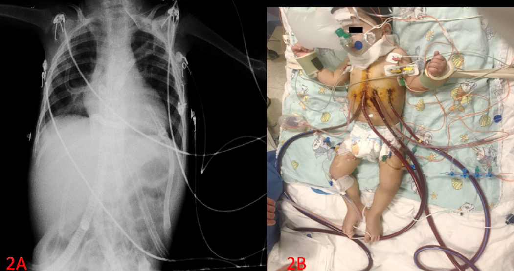
We decided to extubate him due to the clinical state of the lung, and neurologic findings were good despite biventricular dysfunction in the heart. The patient was extubated on the 10th day of ECMO, and subsequently, a non-rebreathing mask was applied (). On the 15th day, the right ventricular support device was removed, and the patient was reintubate before surgery. On the 22nd day after left support device placement, the patient was extubated again (). The left support device was removed on day 26, but the patient was not intubated. On the 34th day of admission, the patient was transferred to the cardiology department with intermittent ventricular tachycardia and a good neurological state.
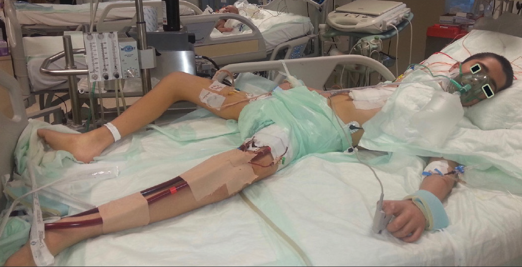
The demographic characteristics, diagnoses, and clinical features of the patients are presented in . Image of the other patients during awake ECMO are shown in respectively.
Table 1. Patients’ demographic, diagnoses and clinical features
Patient 1 | Patient 2 | Patient 3 | Patient 4 | Patient 5 | Patient 6 | |
|---|---|---|---|---|---|---|
Age (y) | 3,5 | 1,5 | 16 | 16 | 13 | 1 |
Gender | Male | Male | Male | Male | Male | Male |
Weight (kg) | 18 | 11 | 40 | 54 | 50 | 7.5 |
Height (cm) | 95 | 80 | 145 | 170 | 170 | 67 |
BSA (m2) | 0.73 | 0.50 | 1.28 | 1.54 | 1.47 | 0.38 |
Primary disease | Necrotizing pneumonia | Myocarditis | Epstein anomaly | Dilated cardiomyopathy- implanted LVAD Heart transplanted | Discordant criss-cross heart | Ventricular septal defect, patent ductus arteriosus |
ECMO indication | Respiratory | Cardiac | Cardiac | Cardiac | Cardiac | Cardiac |
ECMO type | VV | VA | VA | VA | VV, VA-V | VA |
Venous cannula insertion site | RIJV | Right atrium-left atrium (BVAD) | LFV | RFV | RFV, RIJV | RIJV |
Artery cannula insertion site | N | Pulmonary artery-Ascending aorta (BVAD) | LFA | RFA | RFA | Ascendin aorta |
Duration of ECMO (days) | 23 | 28 | 6 (47) | 46 | 28 | 16 |
Complications during ECMO | Bleeding | Clotting in circuit | Limb ischemia | Bleeding, clotting in circuit | Bleeding, Clotting in circuit | Bleeding |
Sedation type during ECMO | Midazolam, fentanyl | Midazolam, fentanyl | Midazolam | Midazolam | Midazolam, fentanyl | Midazolam, fentanyl |
Sedation used during post extubation on ECMO | Fentanyl | Fentanyl | N | N | N | N |
Duration of extubation (days) | 8 | 5 (under BVAD), 4 (under LVAD) | 3 | 6 | 13 | 3 |
Extubation day under ECMO | 5. | 12. | 3. | 4. | 3. | 3. |
Respiratory support while extubated under ECMO | BIPAP | Oxygen mask | BIPAP | BIPAP | Oxygen mask | N-CPAP, BIPAP |
Reintubation during ECMO | Y | Y | Y | Y | Y | N |
Reintubation indication | Respiratory distress | when removing the cannula RVAD | Respiratory distress and hypotension | Respiratory distress and hypotension | Respiratory distress and hypotension | |
On the day after decannulation, it was extubated | Yes | Yes | N | N | N | Y |
Length of duration between decannulation and discharge (days) | 8 | 8 | N | N | N | 95 |
Neurological sequeal | None | None | ||||
Outcome | Survived | Survived | Non-survived | Non-survived | Non-survived | Survived |
BSA: Body surface area, VA: Venoarterial, VV: Venovenous, VA-V: Venoarterio-venous, Y: Yes, N: No, RIJV: Right internal jugular vein, RFA: Right femoral arteria, LFA: Left femoral arteria, LFV: Left femoral vein, BVAD: Biventriculary asist device model, BiPAP: Bilevel positive airway pressure, N-CPAP: Nasal-continuous positive airway pressure
ECMO: Extracorporeal membrane oxygenation
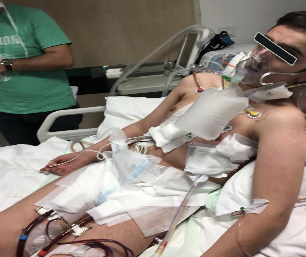
ECMO: Extracorporeal membrane oxygenation
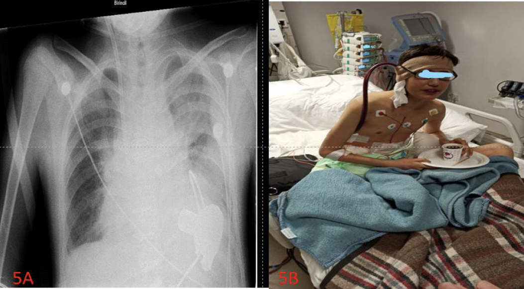
ECMOLVAD: Left ventricular assist device, ECMO: Extracorporeal membrane oxygenation, VV: Venoarteriovenous
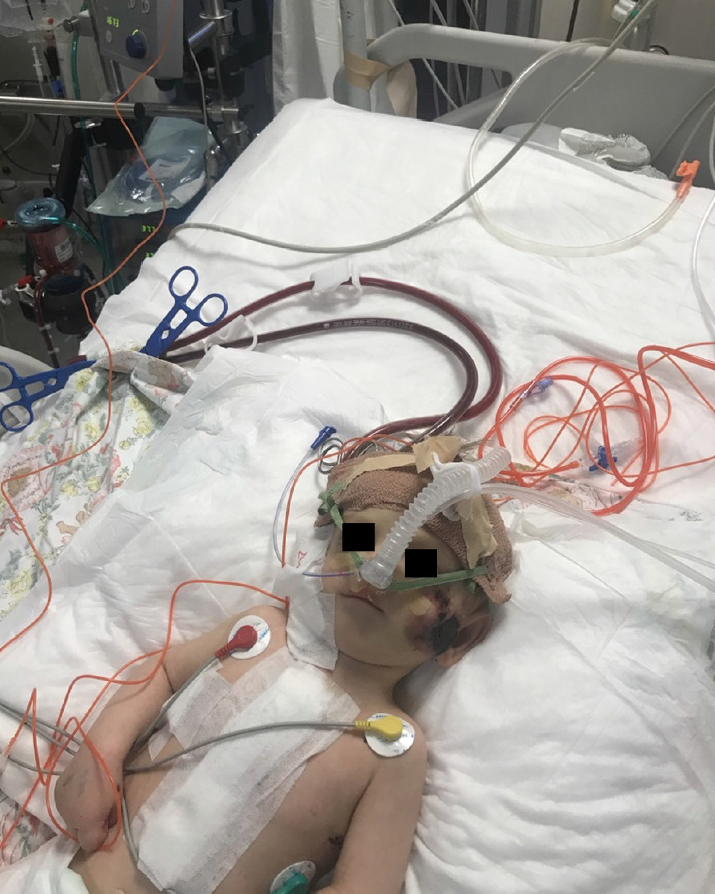
ECMO: Extracorporeal membrane oxygenation, N-CPAP: Nasal-continuous positive airway pressure
Discussion
Patients are often subjected to severe sedation and invasive MV to avoid fatal decannulation during ECMO running., Some centers use extubation during ECMO to completely eliminate barotrauma and volutrauma as a lung rest strategy in adults. Sedation reduces oxygen consumption, thereby improving arterial oxygen transport and organ perfusion, and decreases the irritative effect of the endotracheal tube., However, complications such as sedation habits, dose increase, delirium, or neuromuscular decompression may occur due to sedation. In addition, clinical conditions such as stroke may be masked in patients under sedation., , Benefits include reduced sedation need, increased mobility, and enhanced interaction with the environment. However, the adoption of this application requires careful planning. Patients who are extubated can be reintubate again. In our study on sedation therapy under ECMO, two patients were treated with midazolam and four were treated with midazolam and fentanyl. After extubation while still connected to the ECMO, the dose of sedation treatment was reduced and then stopped.
In addition, awake ECMO is important to assess the clinical and neurological status before transplantation in patients who are candidates for lung transplantation followed by on ECMO. The practice of awake ECMO may strengthen the transplant candidacy by improving the neuromuscular condition of these patients through physiotherapy and advanced nutrition.
A study on awake ECMO in adults was beneficial for pulmonary transplant candidates. In this study, patients intubated during ECMO required longer invasive respiratory support after transplantation and longer intensive care unit stays compared with the awake ECMO group. The 6-month survival rate was higher in the awake ECMO group in this study.
Pediatric awake ECMO was first reported by Anton-Martin
et al.. This study was conducted between 1996 and 2013 with 16 pediatric patients who were extubated while receiving ECMO support. There were 511 patients connected to ECMO. Fourteen of them were connected to ECMO due to respiratory failure and 2 patients for cardiac reasons. Eleven patients had VV ECMO, and 5 patients had VA ECMO. Three patients were reintubate due to arrhythmia and intracranial bleeding. During ECMO, the extubation time was reported as an average of 6 days. Eleven patients who were extubated during ECMO survived. In our study, we reported 6 cases with awake ECMO and followed up 130 pediatric patients who were connected to an ECMO for the same period of time in our PICU.
Similarly, in a study conducted by Schmidt et al. 6 pediatric patients were extubated under ECMO. Six patients were connected to VA ECMO support for cardiac reasons. The number of days extubated under ECMO was 9.5 days, and the mean time to extubation was 78% of the total ECMO time in these patients. The mean duration of ECMO was 17.4 days. One patient was lost during follow-up.
In this study, we presented 6 cases that were extubated while under ECMO. Four of the patients were connected to VA ECMO, and two of them were connected to VV ECMO. Patient 2 was connected to central ECMO, while the others were connected to peripheral ECMO. The durations of our patients’ stays at ECMO were 23, 28, 47, 46, 28, and 16 days, respectively. The patients were extubated while still connected to ECMO on the 5th, 12th, 3rd, 4th, 3rd, and 14th days, respectively. Extubation under ECMO was performed on the 8th, 9th (5 days when the right support device was removed, 4 days when the left support device was removed), 3rd, 6th, 13th, and 3rd days, respectively.
During ECMO, an average extubation time was reported as an average of 7-8 days. Two patients who underwent VV ECMO had the longest extubation time. During awake ECMO, three patients were supported by BIPAP, one patient was supported by N-CPAP, and two patients were given only oxygen supplement. Except for patients 1 and 6, four patients were reintubate for various reasons during ECMO support. Patients 3, 4, and 5 were intubated during decannulation. These patients were reconnected to ECMO because of their worsening status on the same day after decannulation, and three patients were lost despite all interventions during follow-up. Patient 2 was intubated during decannulation, and then patient 2 was extubated. Patient 6 was intubated 29 days after ECMO decannulation. Three patients were extubated during ECMO support and survived.
In our study, ECMO-related complications were observed. ECMO complications are common, including bleeding, stroke, limb ischemia, thrombosis, and infection. Although the most common complications are bleeding (30-40%) and infection (31%), it has been reported that there is at least one major complication in more than half of ECMO patients. In our study, bleeding was observed in four of the patients as an ECMO complication. The ECMO set was changed in 3 patients due to thrombus (changed twice in one patient). None of the patients underwent incidental ECMO decannulation. All patients were treated with heparin during ECMO. The heparin dose was evaluated using activated clotting time and activated partial thromboplastin time tests. CRRT treatment was administered to patients 3, 4, and 5 due to the fluid load under ECMO.
The implementation of awake ECMO requires meticulous team planning and a thorough evaluation of the potential benefits and risks to the patient. To avoid the risks of decannulation, under the supervision of the patient’s parents, nurses, and physicians, the patient’s cannula was also fixed with a coban bandage, and a mesh cap was placed in the exit of the cannula.
Before performing extubation during ECMO, the benefits of extubation should be determined on a patient-based basis. Before this procedure, the expected time to ECMO, possibility of accidental decannulation of the ECMO circuit during extubation, and possibility of emergency reintubation should be considered. This important decision should be made by a multidisciplinary team, and the patient’s family should be informed about this issue.
Conclusion
As a result, the risk of mechanical ventilation-related complications, such as volutrauma and barotrauma, is minimized in patients extubated under ECMO. In addition, sedatives, analgesics, and muscle relax-related complications, such as delirium, muscle weakness, and prolonged ventilation, are reduced via awake ECMO. In patients whose sedation needs are reduced, communication with the environment increases, and clinical conditions such as stroke may be noticed more easily.
Ethics
Ethics Committee Approval: The study was approved by the Institutional Review Board of the Ankara University Faculty of Medicine (approval number: İ06-384-23, 2023/06).
Informed Consent: Voluntary consent was obtained from the families of the patients included in the study.
Footnotes
Authorship Contributions
Concept: E.G., T.K., Design: E.G., E.B., F.K., M.C.S., A.G., T.K., Data Collection or Processing: E.G., E.B., F.K., M.C.S., A.G., T.K., Analysis or Interpretation: E.G., E.B., M.C.S., A.G., T.U., Ö.S.C., E.Ç., M.Ç., Z.E., A.R.A., T.K., Literature Search: E.G., T.U., Ö.S.C., E.Ç., M.Ç., Z.E., A.R.A., T.K., Writing: E.G., T.K.
Conflict of Interest: No conflict of interest was declared by the authors.
Financial Disclosure: The authors declared that this study received no financial support.
Footnotes
Ethics
Ethics Committee Approval: The study was approved by the Institutional Review Board of the Ankara University Faculty of Medicine (approval number: İ06-384-23, 2023/06).
Informed Consent: Voluntary consent was obtained from the families of the patients included in the study.
Footnotes
Authorship Contributions
Concept: E.G., T.K., Design: E.G., E.B., F.K., M.C.S., A.G., T.K., Data Collection or Processing: E.G., E.B., F.K., M.C.S., A.G., T.K., Analysis or Interpretation: E.G., E.B., M.C.S., A.G., T.U., Ö.S.C., E.Ç., M.Ç., Z.E., A.R.A., T.K., Literature Search: E.G., T.U., Ö.S.C., E.Ç., M.Ç., Z.E., A.R.A., T.K., Writing: E.G., T.K.
Conflict of Interest: No conflict of interest was declared by the authors.
Financial Disclosure: The authors declared that this study received no financial support.
References
Schwartz SP, Rehder KJ, Turner DA. Extracorporeal membrane oxygenation survival-more than just decannulation. Pediatr Crit Care Med. 2018;19:905-6.
Schmidt F, Jack T, Sasse M, Kaussen T, Bertram H, et al. “Awake veno-arterial extracorporeal membrane oxygenation” in pediatric cardiogenic shock: A single-center experience. Pediatr Cardiol. 2015;36:1647-56.
Anton-Martin P, Thompson MT, Sheeran PD, Fischer AC, Taylor D, et al. Extubation during pediatric extracorporeal membrane oxygenation: a single-center experience. Pediatr Crit Care Med. 2014;15:861-9.
Ellouze O, Lamirel J, Perrot J, Missaoui A, Daily T, et al. Extubation of patients undergoing extracorporeal life support. A retrospective study. Perfusion. 2019;34:50-7.
Thiagarajan RR. Extracorporeal Membrane oxygenation for cardiac indications in children. Pediatr Crit Care Med. 2016;17(8 Suppl 1):S155-9.
Nosotti M, Rosso L, Tosi D, et al. Extracorporeal membrane oxygenation with spontaneous breathing as a bridge to lung transplantation. Interact Cardiovasc Thorac Surg. 2013;16:55-9.
Mohite PN, Sabashnikov A, Reed A, Saez DG, Patil NP, et al. Extracorporeal Life Support in “Awake” Patients as a Bridge to Lung Transplant. Thorac Cardiovasc Surg. 2015;63:699-705.
Olsson KM, Simon A, Strueber M, Hadem J, Wiesner O, et al. Extracorporeal membrane oxygenation in nonintubated patients as bridge to lung transplantation. Am J Transplant. 2010;10:2173-8.
Jenks CL, Tweed J, Gigli KH, Venkataraman R, Raman L. An international survey on ventilator practices among extracorporeal membrane oxygenation centers. ASAIO J. 2017;63:787-792.
Argent AC. Sedating children on extracorporeal membrane oxygenation: achieving more with less. Crit Care Med. 2017;45:1794-6.
Dalton HJ. Extubation and extracorporeal membrane oxygenation: what a difference a decade makes!! Pediatr Crit Care Med. 2014;15:907-9.
Costa J, Dirnberger DR, Froehlich CD, Beaty CD, Priest MA, et al. Awake Neonatal extracorporeal membrane oxygenation. ASAIO J. 2020;66:e70-3.
Fuehner T, Kuehn C, Hadem J, Wiesner O, Gottlieb J, et al. Extracorporeal membrane oxygenation in awake patients as bridge to lung transplantation. Am J Respir Crit Care Med. 2012;185:763-8.
Mosier JM, Kelsey M, Raz Y, Gunnerson KJ, Meyer R, et al. Extracorporeal membrane oxygenation (ECMO) for critically ill adults in the emergency department: history, current applications, and future directions. Crit Care. 2015;19:431.